
![23 year old with head injury since age 2. Seizures and right hemiparesis since age 10. [Neuroradiology] [CT] [MR]](https://lemmy.world/pictrs/image/39f80f3e-fdaf-4c12-88d7-55e10a61f676.jpeg)
[Left]: Head CT shows left hemispheric volume loss. The injury happened early enough that even the skull is smaller on that side.
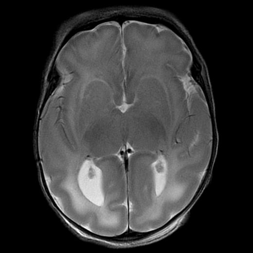
[Right]: Brain MRI shows the severe left hemispheric atrophy. Some of the brain gyri have bulbous ends and a thin neck, resembling mushrooms, a shape called ulegyria and consequence of the brain atrophy. The left lateral ventricle is mildly enlarged due to the atrophied brain.
Just a standard T2 sequence.
FLAIR would have dark CSF, since that’s what FLAIR is designed to suppress - CSF.
MPRAGE is a T1 sequence that’s usually done with contrast.
Got it, thanks for clarifying 👍